
Developmental Biology
Biology

Carl-Philipp Heisenberg
Professor
1 Career
- Since 2010 Professor, Institute of Science and Technology Austria (ISTA)
- 2000 – 2010 Group Leader, Max-Planck-Institute for Molecular Cell Biology and Genetics, Dresden, Germany
- 1996 – 2000 Postdoc, University College London, London, UK
- 1992 – 1996 PhD, Max-Planck-Institute for Developmental Biology, Tübingen, Germany
1.1 Selected Distinctions
- 2019 Carus Medal, German National Academy of Sciences Leopoldina
- 2018 Member of the Board of Reviewing Editors at SCIENCE
- 2017 Lower Austrian Science Award
- 2016 Member of EMBO
- 2015 Member of the German National Academy of Sciences Leopoldina
1.2 Selected Grants
- 2025 – 2030 NOMIS Foundation Research Grant
- 2022 – 2025 HFSP Research Grant
- 2017 – 2022 ERC Advanced Grant
1.3 Tech Transfer (Patents, Spin-Offs)
N/A
1.4 Selected Publications
- Mishra N, Yuting IL, Hannezo É, Heisenberg CP. Geomtry-driven asymmetric cell divisions pattern cell cyles and zygotic genome activation in the zebrafish embryo. Nat Phys. 2026;22, 139-150.
- Caballero-Mancebo S, Shinde R, Bolger-Munro M, Peruzzo M, Szep G, Steccari I, Labrousse-Arias D, Zheden V, Merrin J, Callan-Jones A, Voituriez R, Heisenberg CP. Friction forces determine cytoplasmic reorganization and shape changes of ascidian oocytes upon fertilization. Nat Phys. 2024;20(2):310-321.
- Pinheiro D, Kardos R, Hannezo É, Heisenberg CP. Morphogen gradient orchestrates pattern-preserving tissue morphogenesis via motility-driven unjamming. Nat Phys. 2022;18, 1482–1493.
- Petridou NI, Corominas-Murtra B, Heisenberg CP*, Hannezo E*. Rigidity percolation uncovers a structural basis for tissue phase transitions. Cell. 2021 Apr 1;184(7):1914-1928. *Co-corresponding authors
- Schwayer C, Shamipour S, Pranjic-Ferscha K, Schauer A, Balda M, Tada M, Matter K, Heisenberg CP. Mechanosensation of tight junctions by ZO-1 phase separation and flow. Cell. 2019 Oct 31;179(4):937-952.
2 Group Members
- Laura Hofmann, PhD Student
- Michaela Jovic, PhD Student
- Alena Kizenko, PhD Student
- Roksolana Kobylinska, PhD Student
- Prijwal Patil, PhD Student
- Xin Tong, PhD Student
- Viraj Doddihal, Postdoc
- Dipanjan Ghosh, Postdoc
- Lia Heinemann-Yerushalmi, Postdoc
- Naoya Hino, Postdoc
- Tushna Kapoor, Postdoc
- Long Li, Postdoc
- Carolina Santos Fernandes Lasbarreres Camelo, Postdoc
- Zachary Sun, Postdoc
- Kornelija Pranjic-Ferscha, Research Technician
- Irene Steccari, Research Technician
3 Research
Organismal development emerges from a finely tuned interplay between cell fate specification and morphogenesis- two processes that together transform genetic information into living form. Over the past decades, remarkable progress has been made in uncovering the genetic programs that govern cell fate and patterning. Yet, a fundamental question remains largely unresolved: how do developing organisms physically acquire their characteristic shapes? The molecular, cellular, and biophysical mechanisms driving this transformation are still only beginning to be understood.
For more than 25 years, our research has tackled this challenge through a truly interdisciplinary lens, bridging molecular biology, cell biology, and biophysics. This approach has enabled us to uncover fundamental mechanochemical feedback loops that orchestrate organismal patterning and morphogenesis. We have been at the forefront of redefining how cell adhesion, fate specification, and tissue morphogenesis interact – revealing a dynamic reciprocity in which mechanical forces shape cell adhesion, while adhesion-dependent fate decisions, in turn, remodel cellular mechanics.
In addition, our work has opened new perspectives on the role of cell division in shaping living systems. We have elucidated how cell divisions are executed at the cellular and biophysical level and uncovered their critical contributions to the emergence of form during development.
A defining strength of our research is the seamless integration of experiment and theory, allowing us to connect insights across scales – from molecules to tissues to whole organisms. By leveraging complementary model systems – zebrafish, a key vertebrate model, and ascidians, the closest invertebrate relatives of vertebrates – we are uniquely positioned to uncover evolutionarily conserved principles that lie at the heart of organismal development and morphogenesis.
3.1 Current Scientific Projects/Directions
- The mechanochemical basis of incomplete (meroblastic) cleavage divisions on zebrafish
- Evolution of intermediate filaments and cadherins, and their function in embryo morphogenesis
- The role of environmental oxygen and osmolarity in zebrafish embryo development
- Cytoplasmic organization during vertebrate and invertebrate oogenesis
3.2 Research in Focus
The role of early cleavage divisions in metazoan development is only beginning to come into focus, revealing unexpected layers of regulation at the very onset of life. By combining quantitative experiments with theoretical approaches, we have uncovered a fundamental principle: in zebrafish embryos, the pattern of early cleavage divisions precisely dictates when and where zygotic gene transcription is initiated.
At the mechanistic level, we showed that these cleavage divisions control zygotic genome activation by modulating the nuclear-to-cytoplasmic volume ratio. This ratio is not fixed, but emerges dynamically as successive divisions partition the maternal cytoplasm – often asymmetrically – into daughter cells. Crucially, this process is governed by the geometry of the embryo itself, linking physical constraints directly to gene regulatory control.
In parallel, we investigated the necessity of cytokinesis during these early divisions. Strikingly, we found that abolishing cytokinesis – while leaving nuclear division (karyokinesis) intact – does not impair the cytoplasm’s ability to organize into discrete, cell-like compartments that closely resemble those in normal embryos.
These findings point to a profound and provocative conclusion: canonical cytokinesis may not be essential for early embryonic cleavage. Instead, the fertilized egg appears to possess an intrinsic, self-organizing capacity to partition itself into progressively smaller functional units. This reveals a previously unappreciated robustness and autonomy in early developmental systems, where cellular architecture can emerge from the collective dynamics of the cytoplasm itself.
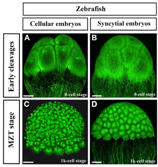
cell-like compartments, which undergo consecutive rounds of divisions that emulate early cleavage divisions in intact embryos. (Heisenberg Lab, unpublished)
(A,B) Exemplary fluorescent confocal images of microtubule organization in cellular and syncytial zebrafish embryos at early cleavage stages (8-cell stage).
(C,D) Exemplary fluorescent confocal images of microtubule organization in cellular and synctial zebrafish embryos at late cleavage stages when maternal-to-zygotic transitions (MZT) occurs (1k-cell stage). Note that syncytial embryos look similar to cellular embryos at early but not late cleavage stages. For visualizing the microtubule cytoskeleton, zebrafish Tg(Xla.Eef1a1:dclk2a-GFP) embryos were used. For inhibiting cytokinesis, embryos were treated with the Aurora Kinase B inhibitor AZD 1152 at 75 μM for 15 minutes at the onset of the first cleavage. Scale bars, 50 μm.

