
Molecular Neuroscience
Neuroscience

Ryuichi Shigemoto
Professor
1 Career
- Since 2013 Professor, Institute of Science and Technology Austria (ISTA)
- 1998 – 2014 Professor, National Institute for Physiological Sciences (NIPS), Okazaki, Japan
- 1989 – 1998 Assistant professor, Kyoto University, Kyoto, Japan
- 1995 PhD, Kyoto University, Kyoto, Japan
1.1 Selected Distinctions
- 2022 Membership Leopoldina
- 2017 Membership Academia Europaea
- 2000 ISI Citation Laureate Award
1.2 Selected Grants
- 2016 – 2021 ERC Advanced grant (694539), SINCHAIS. Awarded to RG: € 2,495,000.
- 2023 – 2025 FWF-ANR grant (I6166-B), PLASTICAZ. Awarded overall: € 696,853; awarded to RG: € 399,000.
- 2025 – 2027 FWF PI grant (PAT5720324), Awarded to RG: € 444,896.74.
1.3 Tech Transfer (Patents, Spin-Offs)
N/A
1.4 Selected Publications
- S. Tabata et al., “Electron Microscopic Detection of Single Membrane Proteins by a Specific Chemical Labeling,” iScience, vol. 22:256-268, 2019, DOI: 10.1016/j.isci.2019.11.025.
- F. Fredes et al., “Ventro-dorsal hippocampal pathway gates novelty-induced contextual memory formation,” Current Biology, vol. 31, 25-38.e5. 2021, DOI: 10.1016/j.cub.2020.09.074.
- P. Bhandari et al., “GABAB receptor auxiliary subunits modulate Cav2.3-mediated release from medial habenula terminals,” eLife, vol. 10:e68274, 2021, DOI: 10.7554/eLife.68274.
- K. Eguchi et al., “Nanoscale Phosphoinositide Distribution on Cell Membranes of Mouse Cerebellar Neurons,” Journal of Neuroscience, vol. 43:4197-4216, 2023, DOI: 10.1523/JNEUROSCI.1514-22.2023.
- P. Koppensteiner et al., “GABAB Receptors Induce Phasic Release from Medial Habenula Terminals through Activity-Dependent Recruitment of Release-Ready Vesicles,” Proceedings of the National Academy of Sciences, vol. 121:e2301449121, 2024, DOI: 10.1073/pnas.2301449121.
2 Group Members
- Abdylla Parahadov, PhD Student since 2021
- Utsa Roy, PhD Student since 2023
- Hana Štefaničková, PhD Student since 2020
- Naoki Zenmyo, Postdoc since 2025
- Pradeep Bhandari, Research Technician since 2024
- Elodie Le Monnier, Research Technician since 2014
- Jacqueline-Claire Montanaro-Punzengruber, Research Technician since 2016
- Elena Säckl, Research Technician since 2016
3 Research
My major goals are to elucidate mechanisms of short- and long-term memory formation in the hippocampus, cerebellum, and medial habenula (MHb) to interpeduncular nucleus (IPN) pathway. We discovered plastic changes of receptor and synapse numbers in physiological learning using SDS-digested freeze-fracture replica labeling (SDS-FRL). These results demonstrate distinct engrams for short- and long-term memory during motor learning (Wang et al., 2014). We also work on the mechanisms of emotional memory formation using the fear conditioning. We discovered that mossy cells in the ventral hippocampus play a critical role in conveying novelty signals to the dorsal hippocampus (Fredes et al., 2021). Another important target is the left-right asymmetry of the brain. Using quantitative SDS-FRL and electrophysiological recording, we discovered that CA1 pyramidal cell synapses have 50% differences in sizes and densities of glutamate receptor subunits between those receiving inputs from the left CA3 and right CA3 (Kawakami et al., 2003; Shinohara et al., 2008). Recently, we discovered another input-side dependent asymmetry in the MHb-IPN pathway (Onal et al., 2026). These scientific achievements depend on electrophysiology and quantitative EM labeling in specific neuronal compartments in the brain. To increase the sensitivity and resolution of the EM labeling, we are also working on development of chemical labeling methods in collaboration with organic chemists and material scientists.
3.1 Current Scientific Projects/Directions
- Left-right asymmetry of brain circuits and functions
- Mechanisms of synaptic plasticity in physiological learning
- Nanoscale physiology of vesicle exocytosis and endocytosis
- Pathophysiology of autoimmune encephalitis
- Development of chemical and proximity labeling using reactive tag systems
3.2 Research in Focus
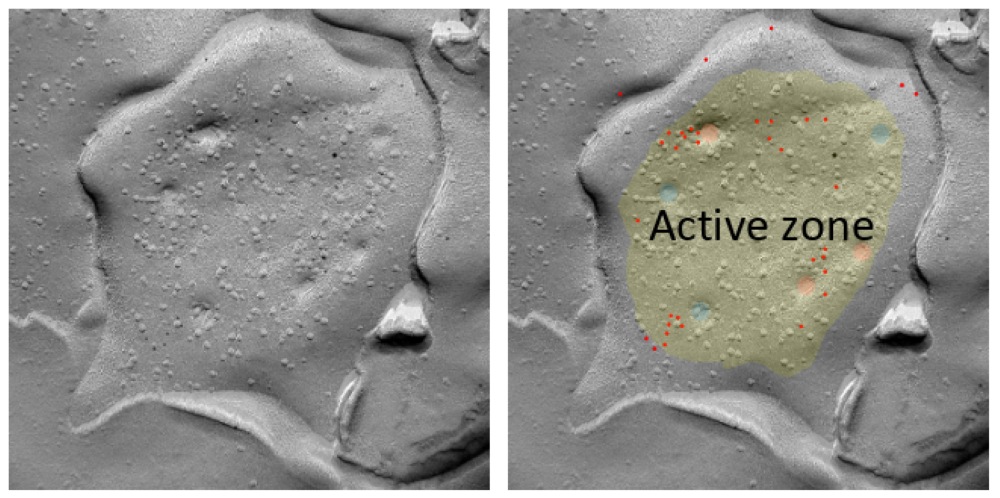
Animals respond rapidly to stimuli because their neurons in the brain are fine-tuned to transmit information within milliseconds of an electrical signal. To enable this, neurons form close physical connections at specialized membrane domains, called synapses. At the synapse, the presynaptic side is packed with many synaptic vesicles (SV) that contain neurotransmitters, for example, glutamate in excitatory axon terminals. These SVs fuse with the presynaptic membrane and release their contents into the synaptic cleft, a phenomenon called exocytosis, to activate the postsynaptic target neuron. The exocytosis occurs within milliseconds of a stimulus, presenting a major challenge for studying activity-dependent structural and molecular changes during neurotransmitter release. In our “Flash & Freeze-fracture replica labeling” technique, we combine optogenetic stimulation of neurons with timed high-pressure freezing, followed by freeze-fracture replica immunolabeling and transmission electron microscopy. Our method captures structural and molecular dynamics snapshots during neurotransmission in millisecond temporal and nanometer spatial resolutions. This work was published in Proceedings of the National Academy of Science, USA: P. Koppensteiner et al., vol. 121:e2301449121, 2024.

