October 11, 2012
An extra dimension in gastrulation
Science issue focusing on "Forces in Development" features two research papers by Carl-Philipp Heisenberg

An issue of Science presenting a collection of articles focusing on “Forces in Development” contains two research papers by the group of Carl-Philipp Heisenberg of the Institute of Science and Technology (IST) Austria, giving new insights into the forces governing gastrulation movements in zebrafish. The first paper, on the role of cortex tension in cell sorting, was already released online at Science Express on August 23. The second paper, looking into contractility in zebrafish epiboly, appears in print for the first time today. In this paper, published together with the group of Stephan Grill at the Max-Planck-Institute of Molecular Cell Biology and Genetics (MPI-CBG) in Dresden, the group uncovers an extra dimension to contractility driving zebrafish gastrulation.
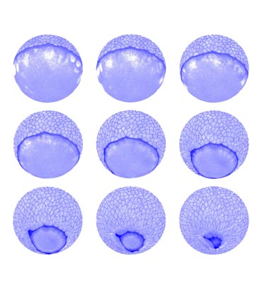
The three embryonic germ layers – ectoderm, mesoderm and endoderm – form during gastrulation. Throughout this process, the shape of the embryo resembles a ball-like sphere. At the onset of gastrulation, the top one third of this sphere is covered by a skin-like layer, called the enveloping cell layer (EVL). In a process called EVL epiboly, this viscoelastic layer will come to cover the entire embryo to provide protection. “You can picture this as a swimming cap that you pull over your head,” says Carl-Philipp Heisenberg. The rim of this cap itself is contractile, and is thought to provide the force that is necessary to stretch the enveloping cell layer over the spherical embryo. Previously, it was believed that the contractile rim acts like a simple purse-string, contracting around the circumference of the spherical embryo. This circumferential contraction would pull the cap over the remaining part of the embryo, once the EVL has passed the sphere’s equator.
In their paper, the group of researchers around Carl-Philipp Heisenberg and Stephan Grill decided to investigate the forces required for EVL epiboly in zebrafish embryos. They show that the rim of the EVL is indeed contractile due to an accumulation of actin and myosin proteins in a ring-like structure at the rim, which is therefore called actomyosin ring. Actin and myosin are components of the cell’s skeleton, where actin forms networks that can be constricted by the motor protein myosin, analogous to muscle contraction. Disruption of this actomyosin ring causes delays in the epiboly process, demonstrating that actomyosin contraction is required for the process.
To their surprise, the researchers also detected significant forces acting in a perpendicular direction, along the width of the actomyosin ring. These experimental results clearly challenged the view of the actomyosin ring being a simple purse-string, solely constricting around its circumference. Indeed, high-resolution imaging of the actomyosin distribution revealed the actomyosin ring to contract not only along its circumference but also along its width. Contraction of the actomyosin ring along its width will lead to a force directly pulling the enveloping cell layer over the sphere, when resisted by some friction to the cytoplasm surrounding the actomyosin ring. This, so Carl-Philipp Heisenberg, “adds – if you wish – an extra dimension to actomyosin ring contraction”.


