April 28, 2014
Watching proteins at work
IST Austria Professor Ryuichi Shigemoto set to revolutionize electron microscopy imaging in the nanometer world • Development supported by Austrian science fund FWF • Method to contribute to EU Human Brain Project and US NIH grant

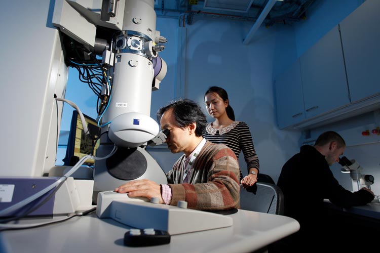
Protein mobility, RNA dynamics, vesicle transport – the Green Fluorescent Protein (GFP) revolutionized cell biology, and the way researchers study the micrometer world. Ryuichi Shigemoto, Professor at the Institute of Science and Technology Austria (IST Austria), seeks a similar revolution for electron microscopy and the nanometer world. In three grants, he and his team intend to develop new methods for electron microscopy with which scientists can study proteins at an unprecedented resolution, employ existing expertise to provide fundamental data for the human brain project, and share methods with international collaborators.
To function, proteins act with other proteins and form stable complexes like ion channels. Currently, researchers can only use biochemical or fluorescent methods to study the composition of proteins and protein complexes. There is as yet no way to see the composition of a single complex in situ in the tissue it acts in. In a stand-alone project funded by the FWF with € 280’000, Ryuichi Shigemoto strives to develop a new method of visualizing single proteins and even subunits of ion channels by electron microscopy (EM). Ryuichi Shigemoto and his colleague Akio Ojida, Professor of Bio-Analytical Chemistry at Kyushu University (Japan), base their method on similar principles as GFP-tagging. A small tag is added to proteins using a knock-in method, so that when the gene coding for the protein of interest is expressed, a small tag is added to this protein. Proteins with a GFP-tag are detected as GFP is fluorescent. In EM, an electron beam scans a sample, and a metal particle that scatters electrons is used to visualize a target.
Shigemoto and Ojida will synthesize chemical probes that covalently attach metal particles to the protein tag. Through the direct labeling of target proteins, this method has a resolution ten times higher than conventional EM methods. Conventional methods use antibodies to add gold particles, and target proteins are distinguished with a 20-30 nanometer resolution. As ion channels are 10 nm large, the subunits forming the channel cannot be seen individually. In comparison, a human hair is 60’000 nm wide.
The new method’s resolution will distinguish molecules that are only 2-3 nanometers apart from each other, giving qualitatively new information on proteins and protein complexes. Similar to the many colors of fluorescent tags, developing a range of tags and probes will allow researchers to look at different proteins in a protein complex at the same time. The Shigemoto lab will use this method to precisely localize ion channels in neurons, analyze the subunits that make up ion channels, and count their absolute numbers. This data is fundamental for analyzing how neurons integrate signals and compute outcomes. However, the new method will find many applications not only in neuroscience, but also in other fields of biology for which visualizing working protein complexes is essential.
With his expertise in EM visualization methods, Ryuichi Shigemoto now also contributes a fundamental “channel-omics” study to the Human Brain Project. Shigemoto joined the HBP, succeeding in a call as one of 22 new project partners selected from 350 proposals. Receptors and ion channels mediate how neurons receive signals, integrate incoming signals and generate output signals. To understand how the brain functions, researchers need to know precisely where receptors and channels are located in neurons. However, their localization and organization in three dimensions is mostly unknown.
As part of the HBP, Ryuichi Shigemoto and his team study the quantitative distribution of these key proteins, asking exactly where specific receptors and channels are in a neuron, how many of them there are, and how this differs between types of neurons in the cortex. In a first step, the researchers focus on glutamate receptors, which play a major role in the transmission of excitatory signals, and on voltage-gated calcium channels, major players in the integration of dendritic excitability and release of neurotransmitters. The researchers will expand their analysis to a larger number of channels for an “omics”-type of understanding of ion channels, building up a “channel-omics” for cerebral neurons in the future.
This knowledge feeds into the entire HBP, as the location and quantification of key molecules on neuronal membranes provide fundamental information for the functional analysis and modelling of the brain. The number of channels and channel types in a neuron change the properties of integration and excitability, both key parameters for neuronal simulation. Ryuichi Shigemoto’s data will therefore be integrated with data generated by The Neuroinformatics Platform of the HBP, and provide input for the brain models of the The Brain Simulation Platform.
To round it off, Shigemoto will be supporting collaborator Maria Rubio, Professor at the University of Pittsburgh to utilize high resolution EM visualization methods as part of a National Institute of Health (NIH) grant. Rubio works on glutamate receptors subunits in auditory nuclei.


