August 31, 2023
A Look Into the Brain’s Architecture
Scientists at ISTA present new microscopic technique that reveals the brain in unprecedented detail
The Danzl group at the Institute of Science and Technology Austria (ISTA) presents a new feline-inspired microscopic technique. With “CATS”, scientists are now able to visualize the brain’s architecture in stunning detail across spatial scales and get insights into how it is altered in human brain disease. The new study is published today in Nature Biotechnology.
The human brain is an exceedingly complex organ. Billions of nerve cells are connected by an even larger number of synapses, forming a network that processes our perceptions, orchestrates body function, and allows us to move, think, and feel.
The brain’s highly sophisticated organization spans the size scales of different brain regions working together over centimeters down to the individual cells and the trillions of synapses that connect each nerve cell to others. At these synapses, the minuscule protein machineries that transmit the information are arranged within a space of tens of nanometers. Light microscopy is a powerful visualization tool; however, it lacks the resolving power to decode the fine-grained cellular architecture of brain tissue and, even more so, the synaptic connections between cells.
Through a novel method, presented today in Nature Biotechnology, a new avenue has emerged. The Danzl group and collaborators at the Institute of Science and Technology Austria (ISTA) established an innovative technique, dubbed CATS (Comprehensive Analysis of Tissues across Scales), which enables scientists to examine brain tissue architecture and thereby get a detailed look into the entire cellular composition of the tissue. This advancement facilitates a better understanding of how cells and their synapses relate to each other and provides cellular context for specific molecules.
Reconstructing single synapses in 3D
To visualize cells with microscopic techniques, the cells first need to be labeled. Common labeling approaches, including the use of fluorescent dyes, however, remained limited to a few highlighted cells or molecules devoid of their context.
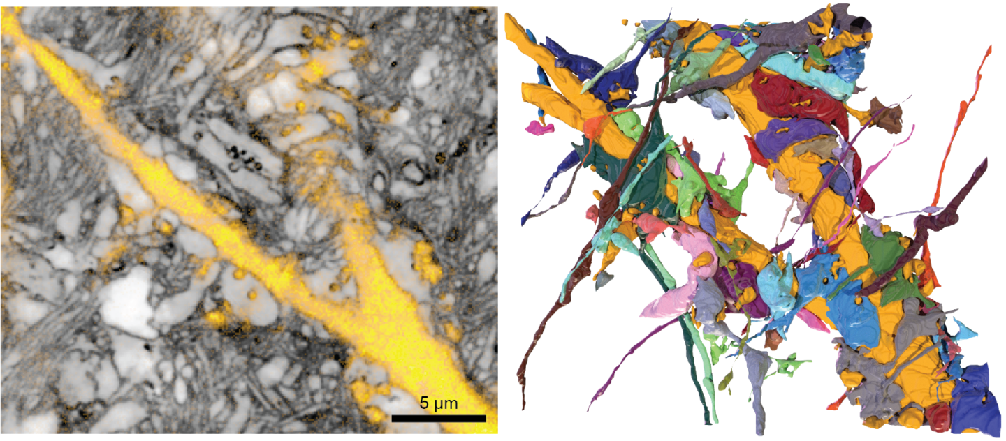
For their novel technique, the researchers devised a strategy to label the cell surfaces and spaces around cells in a way that is compatible with chemically conserving or “fixing” the tissue. Here, the cells themselves appear as negative shadows. Employing “super-resolution” light microscopy techniques—an enhanced light microscopy technique—for readout then allows to decode the tissue architecture at a much finer resolution than a conventional light microscope would be capable of. To quantify the images, the researchers integrated labeling and super-resolution imaging with advanced analysis, capitalizing on the ongoing revolution in deep learning technology.
With CATS, scientists can now relate the location of molecular machineries to cells and synapses, reconstruct single synapses in 3D, and determine which of the neighboring cellular structures form synaptic connections with a given cell.
“We’re excited about the CATS technology, as it adds crucial information that is missing in light microscopy analysis. CATS visualizes brain tissue architecture in its entirety, rather than focusing just on a few cells. It locates cells and specific molecules in the micro- and nanoscale context, allowing us to decode how the functional components of the tissue relate to each other,” explains Johann Danzl.
Achieving scientific breakthroughs through interdisciplinarity
The CATS project combined expertise from vastly different areas. The Danzl group collaborated with neuroscientists at ISTA (the Siegert, Novarino & Jonas groups) to integrate diverse brain tissue preparations and functional measurements on neuronal activity and with neurosurgeons and neuropathologists from the Medical University Vienna, to apply the technology to specimens from human brain surgeries and archival pathology samples. This enabled the researchers to analyze the deranged tissue architecture in human brain disease with greater precision.
on the project,”For me, it was a huge drive to be part of this process,” says first author and previous PhD student in the Danzl group, Julia Michalska. “I am proud that we were able to bring together such a distinguished, interdisciplinary group of researchers for this project and that we have been able to contribute this tool to the scientific community, which hopefully will be instrumental in the years to come.”
Publication
J. M. Michalska, J. Lyudchik, P. Velicky, H. Štefaničková, J. F. Watson, A. Cenameri, C. Sommer, N. Amberg, A. Venturino, K. Roessler, T. Czech, R. Höftberger, S. Siegert, G. Novarino, P. Jonas & J. G. Danzl. 2023. Imaging brain tissue architecture across millimeter to nanometer scales. Nature Biotechnology. DOI: 10.1038/s41587-023-01911-8
Full text link (open access): https://www.nature.com/articles/s41587-023-01911-8
Funding information
The ISTA project was funded by the following sources:
Austrian Science Fund (FWF) grant I3600-B27 (J.G.D.); Austrian Science Fund (FWF) grant DK W1232 (J.G.D. and J.M.M.); Austrian Science Fund (FWF) grant Z 312-B27, Wittgenstein award (P.J.); Austrian Science Fund (FWF) projects I4685-B, I6565-B (SYNABS) and DOC 33-B27 (R.H.); Gesellschaft für Forschungsförderung NÖ (NFB) grant LSC18-022 (J.G.D.); European Union’s Horizon 2020 research and innovation programme, European Research Council (ERC) grant 715508 – REVERSEAUTISM (G.N.); European Union’s Horizon 2020 research and innovation programme, European Research Council (ERC) grant 692692 – GIANTSYN (P.J.); Marie Skłodowska-Curie Actions Fellowship GA no. 665385 under the EU Horizon 2020 program (J.M.M. and J.L.); and Marie Skłodowska-Curie Actions Individual Fellowship no. 101026635 under the EU Horizon 2020 program (J.F.W.).
Information on animal studies
In order to better understand fundamental processes, for example, in the fields of neuroscience, immunology, or genetics, the use of animals in research is indispensable. No other methods, such as in silico models, can serve as an alternative. The animals are raised, kept, and treated according to the strict regulations of Austrian law. All animal procedures are approved by the Federal Ministry of Education, Science and Research.
