August 12, 2026
Brain and Organoid Side-by-Side
ISTA researchers develop a new organoid model of the cerebral cortex
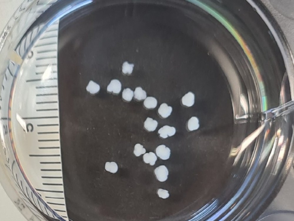
For almost 15 years, researchers have been creating three-dimensional biological microstructures known as organoids—from heart and gut models to brain models. These allow for the study of specific questions about organ development in an isolated system. The Hippenmeyer group at the Institute of Science and Technology Austria (ISTA) now presents its first organoid model of the mouse cerebral cortex in Nature—and thus provides new insights into its development in a Petri dish as well as in living organisms.
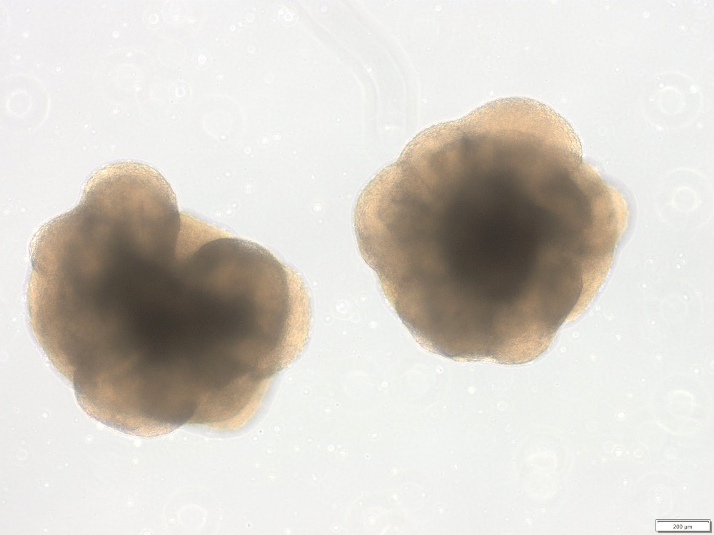
Over a decade ago, scientists redefined how to study organ development by generating a seemingly unremarkable clump of cells. They dubbed them organoids: an organ-like microstructure produced using stem cells. Stem cells are cells that have two important properties: they can make more of themselves and they can divide to produce more specialized cells, like neurons or muscle cells.
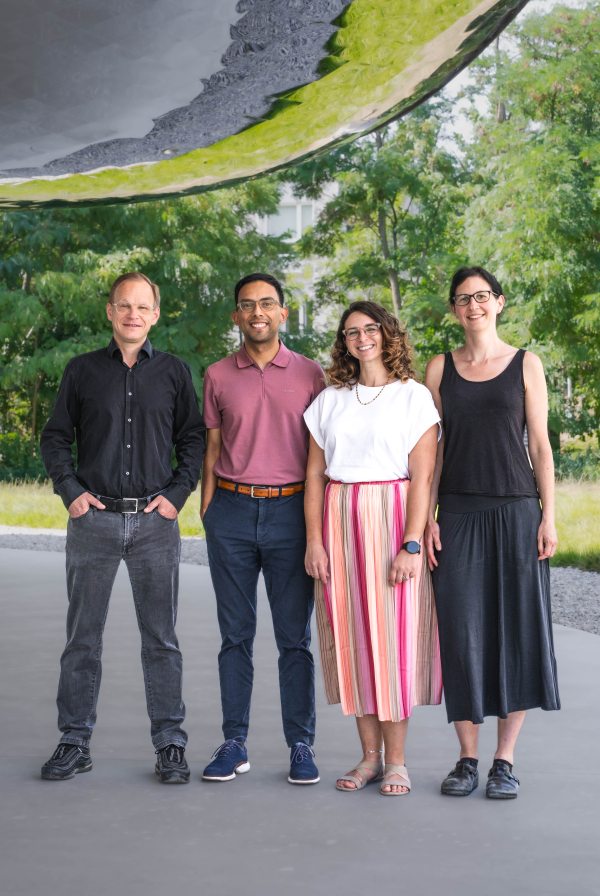
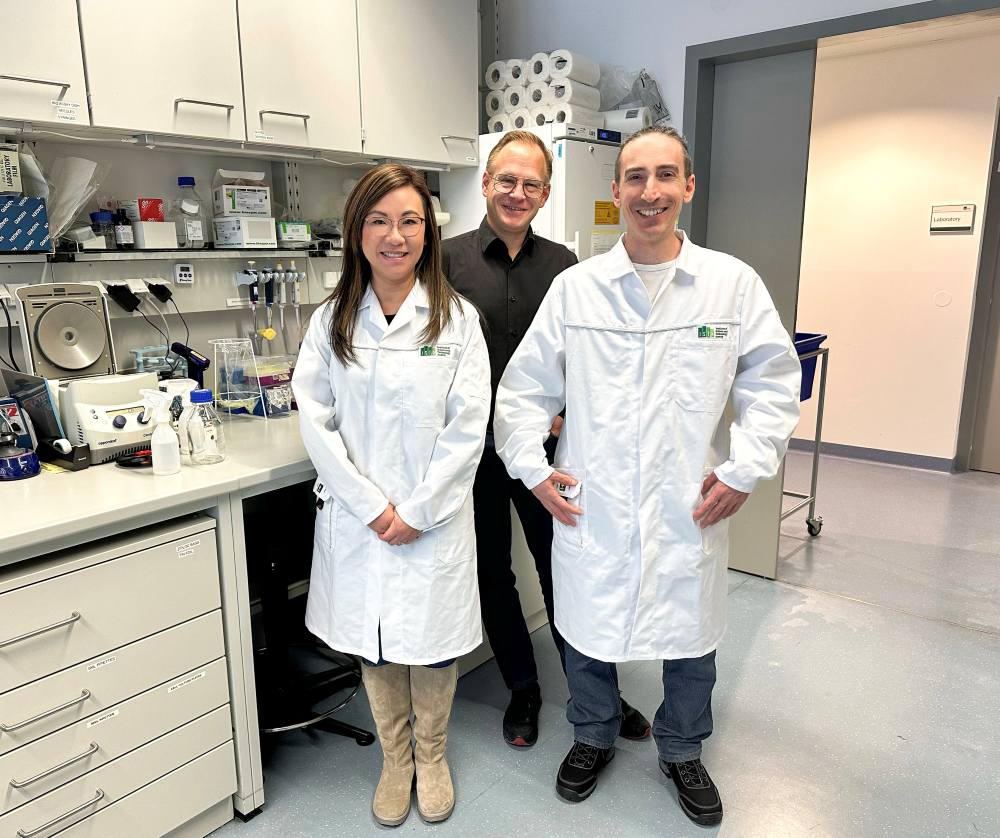
In Nature, the Hippenmeyer group—in particular co-lead authors Melissa Stouffer, Osvaldo Miranda Romero, Florian Pauler, and Fabrizia Pipicelli, along with Carmen Streicher and Giselle Cheung—presents its first organoid model of the mouse cerebral cortex. They discovered critical time windows in development when compared to the real mouse brain.
From stem cells to a correctly-sized brain
“In our lab we study how the brain develops from stem cells,” Hippenmeyer explains. “How a brain reaches the right size, how stem cells know when and into which neurons they should develop, but also what happens when something goes wrong during development or disease—for example, in microcephaly or macrocephaly, where the brain is unusually small or large.”
As in countless laboratories across the world, most of the research conducted in the Hippenmeyer group is done using mice. The team uses mice to study the cerebral cortex—a brain region densely packed with neurons and glial cells, and one that is critical for brain function and cognition.
“About eight years ago, we started looking more closely into organoids,” he continues.
At the time, most organoids were made from human stem cells—which made sense, as they are intended to provide insights into human developmental processes as closely as possible. However, human stem cells are only suitable for genetic experiments to a limited extent and, compared to mice, fewer established genetic tools are available.
Together with Melissa Stouffer, then a postdoctoral researcher, the Hippenmeyer group set out to develop organoids that could be used to closely investigate questions related to the development of the cerebral cortex.
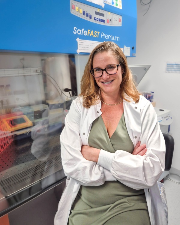
New expertise, new tools, and unprecedented insights at single-cell resolution
Organoid research is complex and requires a particular skillset. Stouffer therefore attended the Brain Organogenesis Workshop at Stanford and immersed herself into the subject.
“That was a game changer,” Stouffer recalls. “It allowed me to appreciate the intricacies of 3D in vitro techniques and the difficulty in working with stem cells. Prior to setting up any specific quantifications in the organoids, I needed to ensure that I had derived high-quality stem cell lines and developed a protocol that was reproducible across batches and cell lines. This groundwork was time-consuming but was the foundation for all subsequent steps with the organoids.”
With this expertise and the right tools, the Hippenmeyer group eventually developed a stable mouse stem cell line. They used this novel line to develop a robust and reproducible mouse cortical organoid protocol.
But how similar are these three-dimensional structures to the real thing?
To understand how close a developing organoid is to the real brain, it makes sense to look at both in the same species—not to compare a mouse brain with a human organoid, but to study both in the mouse model.
The researchers therefore compared specific developmental stages of the mouse brain with those of the organoids. Using single-cell sequencing, they examined which cell types show up in both systems, their relative abundance, and at what point in time they emerge or disappear again. “In the developmental stages we examined, we see very similar cell populations of the mouse brain with those of the organoids,” Hippenmeyer explains. “The molecular programs are similar.”
Similar program, different timing
“Now that we had this rigorous organoid system, we were able to examine even more closely what happens to stem cells during cortical structure development—and compare these processes directly with our in vivo model, the mouse,” Hippenmeyer continues.
Using the MADM (Mosaic Analysis with Double Markers) technique—a unique genetic method that makes it possible to track stem cell division during organogenesis, the phase where organs start to form from the three primary germ layers—the Hippenmeyer group has already produced a clear roadmap of development in the mouse brain at the single progenitor cell level.
In vivo, development follows a temporally linear model. At first, stem cells multiply through symmetric division and then switch to asymmetric division for a short time in order to produce neurons. Only once all neurons are made, glial cells emerge. The individual phases therefore take place sequentially—not at the same time. Alterations to the finely tuned developmental process, for example through genetic mutations, can lead to severe brain malformations, such as microcephaly or macrocephaly.
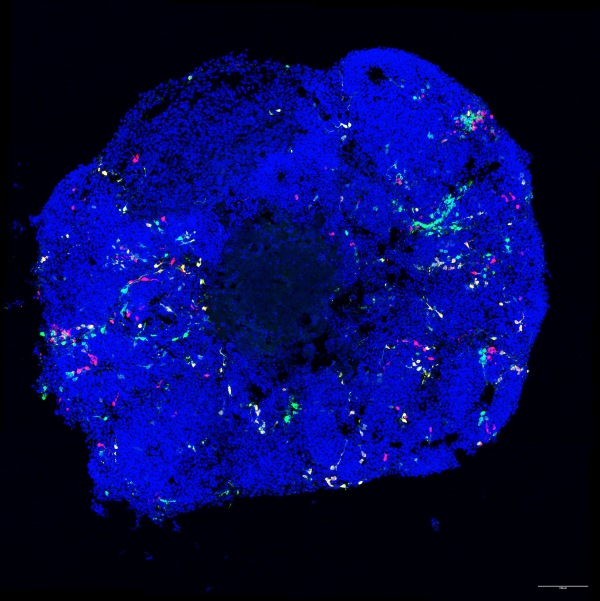
At the macro level, cortical organoids are similar enough to be able to give scientists insights into broad aspects of development. However, stem cells in cortical organoids lack the fine-tuned progression of developmental stages found in the mouse. The same cell types emerge, but the timing of neuronal development is uncoupled in the organoid.
In other words, brain stem cells in the organoid lack a proper sense of time. They keep the hour but not the minutes. One exception is the establishment of glial cells: this process also takes place after neuron formation in the organoid—just as it does in the mouse brain.
Importantly, the revelation of these specific aspects of similarity and divergence from the in vivo condition represents a major conceptual advance for the better understanding of the mechanistic framework.
The researchers suspect that organoids lack certain external signals. The microstructures in Petri dishes form through self-organization, and due to being cultured in the lab, they lack many of the external influences that are present in the living organism.
“In our organoids, this does not seem to work perfectly,” says Hippenmeyer. “The physical force of self-organization alone is apparently not enough. Factors present in in vivo systems are missing—the so-called stem-cell niche.” The stem-cell niche is the specific microenvironment in which stem cells live and are regulated. It includes, for example, neighboring cells, blood vessels, signaling molecules, and growth factors, as well as mechanical signals.
What comes next?
The Hippenmeyer group’s new publication presents, on the one hand, a robust protocol for producing cortical organoids from mouse cells. On the other hand, it also highlights the processes in development which are sensitive to changes to—or a lack of—the stem cell niche.
These findings are important for organoid research. They show how similarly certain developmental processes unfold in organoids and in the mouse brain—and up to what point, based on current knowledge, specific aspects of brain development can be reliably studied in an organoid.
As a next step, the researchers want to try to recreate aspects of the stem-cell niche in the organoids. To do so, they could gradually add factors that are present in the mouse brain and may help restore the temporally linear sequence of development.
Publication:
M. Stouffer*, O. A. Miranda*, F. M. Pauler*, F. Pipicelli*, C. Streicher, G. Cheung & S. Hippenmeyer. 2026. Temporal Uncoupling of Radial Glia Lineage Progression in Cortical Organoid. Nature. DOI: 10.1038/s41586-026-10916-7
*Denotes equal contribution.
Funding information:
This project was supported by funding from the European Commission (IST plus postdoctoral fellowship), by ISTA institutional funds, FWF SFB F78 Neuro Stem Modulation, and by the European Research Council (ERC) under the European Union’s Horizon 2020 research and innovation program (grant agreement 725780 LinPro).
Information on animal studies:
In order to better understand fundamental processes, for example, in the fields of neuroscience, immunology, or genetics, the use of animals in research is indispensable. No other methods, such as in silico models, can serve as an alternative. The animals are raised, kept, and treated according to the strict regulations of Austrian law. All animal procedures are approved by the Federal Ministry of Women, Science, and Research.