Microscopy
2 x Inverted widefield microscope Nikon Eclipse / Ti-2E
- Widefield fluorescent microscope
- Lumencor SpectraX light source
- Motorised stage
- cameras:
- Nikon Eclipse: color (Nikon DS-FI) and monochrom (Hamamatsu EMCCD 9100)
- Nikon Ti-2E: DS-Qi2
- Temperature control chamber
- external phase contrast and DIC imaging optics
- 1 system (Nikon-Ti-2E) with automated experiment design option
Upright LSM 800 confocal microscope

- Laser lines 405, 488, 555, 640 nm
- 2 GaAsP PMT’s for reflected light detection
- PMT for transmitted light detection
- Multiple position imaging + automated experiment design option
- Temperature chamber available
3 x Inverted LSM 800 confocal microscope
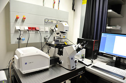
- 3 x Laser lines 405, 488, 555, 640 nm
- 2 setups
- 1 x MA PMT’s for reflected light detection
- 2 x GaAsP PMT’s for reflected light detection
- ESID + LED electronically switchable illumination and detection module
- Multiple position imaging + automated experiment design option
- The 2 systems with GaAsP detectors are equipped with incubation chamber and CO2/N2 mixer
2 x Vertical LSM 800 confocal microscope
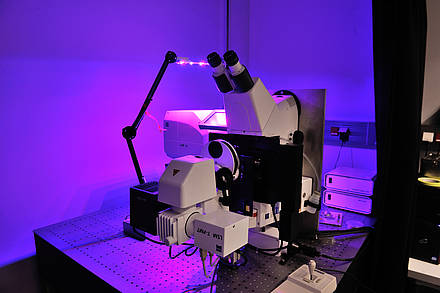
- Microscope is turned at 90° to enable observation of sample in a proper position to gravity
- Laser lines: 405, 488, 555, 640 nm
- 2 MA-PMT’s for reflected light detection
- ESID + LED electronically switchable illumination and detection module
- Multiple position imaging + automated experiment design option
- Rotation stage to change the angle of a sample in relation to gravity
- Custom-programmed ‘root tracker’ software to adjust stage position to the sample movement
Upright LSM 880 confocal microscope with Airyscan
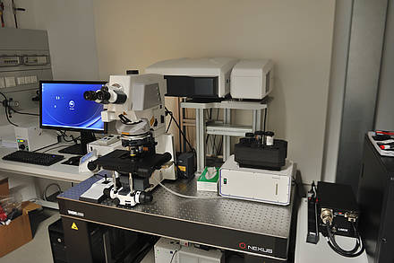
- Laser lines 405, 458, 488, 514, 561, 640 nm
- 2 PMT’s and 1 GaAsp (single channel) detector for reflected light detection
- Airyscan detector
- PMT for transmitted light detection
- Multiple position imaging + automated experiment design option
- Equipped with incubation chamber for temperature and a gas-mixer
Inverted LSM 880 confocal microscope with Airyscan


- Laser lines 405, 458, 488, 514, 561, 640 nm
- 2 PMTs and 1 GaAsp (single channel) detector for reflected light detection
- Airyscan detector
- PMT for transmitted light detection
- Multiple position imaging + automated experiment design option
- Equipped with incubation chamber for temperature and a gas-mixer
Upright SP5 confocal microscope
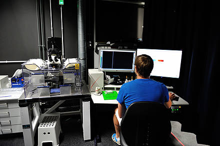

- Laser lines 405, 458, 488, 514, 561, 633
- 5 PMTs with free selection of detection range
- TL detector for transmitted light
- Motorized stage for multiple position imaging
- Temperature-control chamber
Inverted SP5 confocal microscope
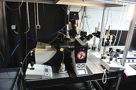

- Laser lines 458, 488, 514, 561, 633
- 3 PMTs and 2 hybrid detectors (Leica HyD) with free selection of detection range
- transmitted light detector
- Tandem scanner for ultrafast imaging
- Heated stage and objective heater
- FRET and FRAP software
- Micromanipulators / microfluidic pump
Upright multiphoton microscope and FLIM Detector
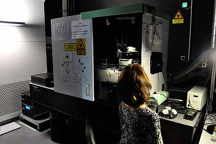

- Upright 2-photon microscope
- Multiphoton imaging with two laser lines (Cameleon Ultra II and OPO compact), wavelength range 780-1300 nm
- Multichannel MP imaging with up to 4 NDD (all GaAsp detectors) simultaneously
- 2 PMTs in a transmitted port, for SHG/THG imaging
- Incubation chamber for temperature control
- Can be equipped with portable PMT based FLIM X16 TCSPC [Time Correlated Single Photon Counting] detector
Inverted multiphoton microscope
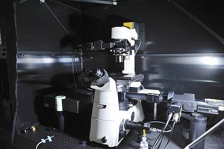

- Inverted 2-photon microscope
- Multiphoton imaging with two laser lines (Cameleon Ultra II and OPO compact), wavelength range 780-1300 nm
- Multichannel MP imaging with up to 3 NDD (all GaAsp detectors) simultaneously
- EOM control of laser power, ROI imaging, linescan
- 1 PMTs in a transmitted port, for SHG/THG imaging
- Tabletop sample incubator and portable CO2 mixer
- Portable PMT based FLIM X16 TCSPC [Time Correlated Single Photon Counting] detector
Spinning disk & laser cutter microscope
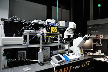

- Inverted Microscope
- Spinning Disk confocal CSU-X1
- 488,561,640 nm laser lines
- Camera: iXon 897
- FRAPPA unit
- Scanning laser microdissection with pulsed 355 nm laser, simultaneous imaging and microdissection possible
- Motorized piezo stage
- stage and objective heating unit
- Micromanipulators / microfluidic pump
Spinning disc and TIRF microscope iMic
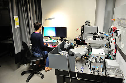

- Inverted Spinning Disc / multipoint TIRF / epifluorescence microscope
- Spinning disc confocal unit: Andromeda
- Spinning disc unit camera: Andor Ixon 885
- Laser lines: 405, 445, 488, 561, 640 nm
- Oligochrome light source for epifluorescence imaging
- Multipoint 360° TIRF (Total Internal reflection)-microscopy with all laser lines, motorized adjustment of TIRF angle calibration
- FRAP with all laser lines
- TIRF/Epifluorescence port is equipped with image splitter (Andor Tucam) with two Ixon 897 cameras
- Motorized stage for multiposition imaging
- Temperature control of a sample with tabletop incubator (incubation chamber coming soon)
TIRF / FRAP microscope
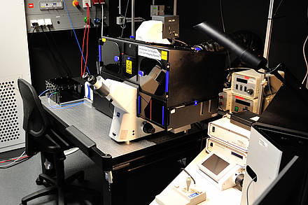

- Automated TIRF/Epifluorescence microscope
- Multipoint 360° TIRF (Total Internal reflection)-microscopy with all laser lines (iLAS2), motorized adjustment of TIRF angle calibration
- Laser lines 405, 488, 561, 640nm
- Lumencor SpectraX engine for epifluorescence imaging
- Equipped with FRAP module for simultaneous and sequential bleaching (405 nm)
- Equipped with climatised chamber- temperature control, CO2 supply
- DIC imaging is possible combined with epifluorescence
OpenSPIM Light Sheet microscope
- 488 nm and 561 nm lasers
- 525/50 BP filter for GFP imaging, dual bandpass filter for green/red imaging
- Camera: Hamamatsu Orca R2
- Temperature controlled sample chamber, peristaltic pump
- multiple sample chambers for different RI / sample embedding media
- OpenSPIM open source platform information website: openspim.org
CellHesion Atomic Force microscope
Probing Atomic Force Microscope with possibility of simultaneous fluorescence imaging
- 2 different scan heads:
- JPK Cellhesion 200: force spectroscopy
- Nanowizard 4: AFM scanning head for contact, tipping or quantitative imaging
- Petri dish heater unit
- High sensitivity cooled Andor camera
- in-house developed top-view optics
VS120 Slide Scanner, Olympus
- Widefield microscope
- Grayscale and colour camera
- Light source: X-Cite Excite lamp
- Filter combinations for DAPI, FITZ, CY3, CY5
- Motorized stage with 5 slide positions
Inverted widefield Olympus IX83 microscope
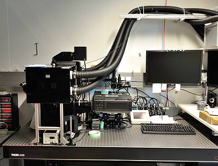

- Widefield fluorescent microscope
- Lumencor SpectraX light source
- Motorised stage
- Hamamatsu Orca Flash 4.0 sCMOS camera
- Temperature control chamber


