May 24, 2021
Defective Gene Slows Down Brain Cells
Scientists at IST Austria discover how a high-risk gene for developing autism spectrum disorder affects brain development. Study published in Nature Communications.
© IST Austria
Although many forms of autism spectrum disorder (ASD) are thought to have genetic causes, the cellular and molecular functions of the identified genes remain unclear. Scientists at the Institute of Science and Technology (IST) Austria studied a high-risk gene and discovered its important role during a critical phase of brain development.
Within the European Union alone, about three million people are affected by an autism spectrum disorder (ASD). Some are only mildly affected and can live independent lives. Others have severe disabilities. What the different forms have in common is difficulty with social interaction and communication, as well as repetitive-stereotypic behaviors. Mutations in a few hundred genes are associated with ASD. One of them is called Cullin 3, and it is a high-risk gene: A mutation of this gene almost certainly leads to a disorder. But how exactly does this gene affect the brain? To learn more about it, Jasmin Morandell and Lena Schwarz, PhD students at Professor Gaia Novarino’s research group, turned to mice whose Cullin 3 gene has been partially deactivated and compared them to their healthy siblings. Their results have just been published in the journal Nature Communications.
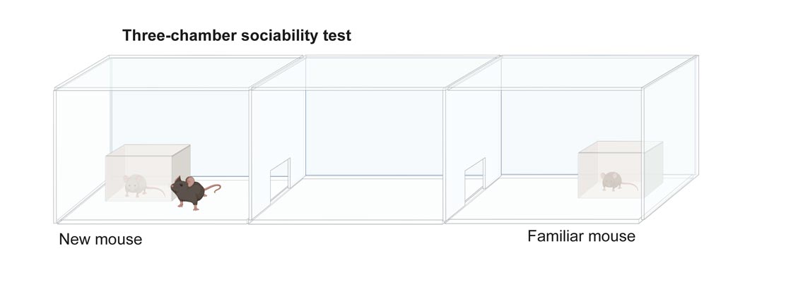
In a series of behavioral and motoric tests, the team wanted to see if the modified mice mimicked some of the characteristics of patients with this form of autism and could therefore be used as model organisms. In one of these tests, the so-called three-chamber sociability test, a mouse could freely explore three adjacent chambers of a box connected by little doors. Now, the scientists put two other mice in the outer boxes: One was already familiar to the studied mouse, the other mouse it had never met. “Healthy mice usually prefer the new over the already familiar mouse,” Jasmin Morandell, co-first author of the study, explains. The mouse with the altered Cullin 3 gene, however, showed no sign of recognition. Furthermore, the mice had motor coordination deficits as well as other ASD-relevant cognitive impairments. With the help of this mouse model, the team was then able to get to the bottom of the mechanisms that bring about these changes.
A Dangerous Accumulation of Proteins
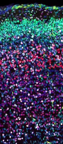
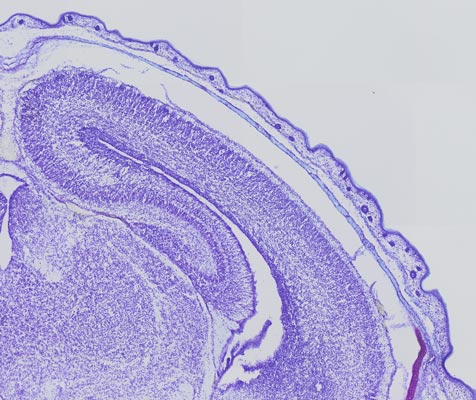
While studying the mouse brain, the researchers noticed a very subtle but consistent change in the position of some brain cells. These so-called neurons or nerve cells originate from a special region in the brain. From there, they migrate toward the uppermost layers until they find their designated place in the cortex. It is a very sensitive process, where even small changes in the speed at which they travel can change the structure of the cortex. By marking the migrating neurons, the scientists could trace their movements. “We could observe migration deficits – the neurons are stranded in the lower cortex layers,” Lena Schwarz, the other co-first author of the study, describes. But why are the cells not moving as they should?
The answer lies in the important role Cullin 3 plays at the end of life of proteins. When their time has come, the gene Cullin 3 tags them for degradation – a process that has to be tightly regulated to prevent proteins from accumulating. To find out, which proteins are misregulated when Cullin 3 is defective, Morandell and Schwarz systematically analyzed the protein composition of the mouse brain. “We were looking at proteins that accumulate in the mutant brain and found a protein called Plastin 3. Then Gaia came across a poster describing the work of IST Austria’s Schur group in the hallway, and we got very excited,” says Jasmin Morandell. “They independently had been working on Plastin 3 as a regulator of cell motility and had complementary results to ours. That’s when we started working together,” Professor Gaia Novarino remembers.
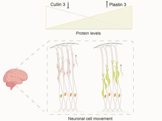
It turned out that the protein Plastin 3, which was previously unknown in the context of neuronal cell migration, actually plays an important role in this process. “If the Cullin 3 gene is deactivated, the Plastin 3 protein accumulates, causing cells to migrate slower and over shorter distances. This is exactly what we saw happening in the cortex of the Cullin 3 mutant mice,” says PhD student Lena Schwarz.
A Risky Pathway?
All this is taking place during a very early stage of brain development around halfway through pregnancy – long before anyone would notice any difference in the fetus. “Determining these critical windows during brain development could be extremely important to fine-tune the treatment of patients with specific forms of ASD,” explains Novarino, who is committed to improving diagnosis and treatment options for people with ASD. “Following up with the research on Plastin 3 could pave the way for some therapeutics. Inhibiting the accumulation of this protein could eventually alleviate some of the symptoms patients have,” Schwarz says.
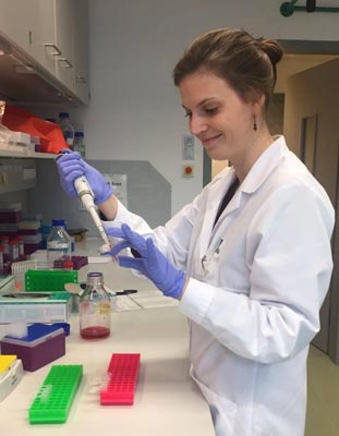
Graduate Jasmin Morandell. © IST Austria 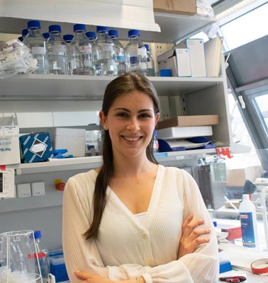
PhD student Lena Schwarz. © IST Austria 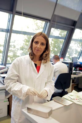
Professor Gaia Novarino. © Gerald Riedler, IST Austria
“We now know that defective Cullin 3 leads to increased levels of Plastin 3. This tight correlation shows that Plastin 3 protein levels may be an important factor for the control of cell-intrinsic movements,” says Jasmin Morandell. She recently graduated and may use her expertise in brain development to study Huntington’s disease. Lena Schwarz will next turn to additional high-risk ASD genes to see how other proteins in the degradation pathway may be linked to ASD. For the present study, the Novarino group joined forces with the Danzl and Schur groups and a colleague from the University of Rome. “Finishing this extensive study in around two and a half years despite the pandemic was only possible with the support from our neighbors at IST Austria,” Novarino praises the multidisciplinarity at the Institute.
Publication
Jasmin Morandell, Lena A. Schwarz et al. 2021. Cul3 regulates cytoskeleton protein homeostasis and cell migration during a critical window of brain development. Nature Communications. DOI: 10.1038/s41467-021-23123-x
Funding information
The IST Austria project part was supported by funding from the European Union’s Horizon 2020 research and innovation program (ERC) and by the Austrian Science Fund (FWF) to Gaia Novarino and to Johann Georg Danzl.
Animal welfare
In order to better understand fundamental processes, for example, in the fields of neuroscience, immunology, or genetics, the use of animals in research is indispensable. No other methods, such as in silico models, can serve as alternatives. The animals were raised, kept, and treated according to the strict regulations of Austrian law.
